
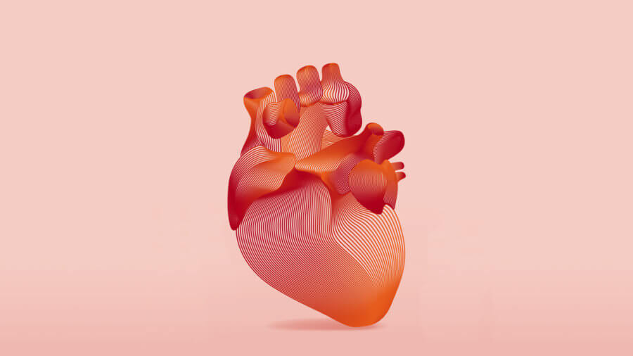
Hearts aren’t meant to be broken. Yet with age, it happens. Even with a healthy diet and exercise, as our age slowly ticks up, so does the risk for clogged arteries, brittle blood vessels, and ultimately heart failure.
Why?
Scientists have long sought to untangle the mystery of how aging links to increased risk of heart disease, a predominant killer of our time. It’s a tough problem: many biological aspects, spanning nature to nurture, can subtly influence heart health. To untangle the mystery, some experiments have lasted over half a century and scaled to hundreds of thousands of people.
The good news? We’ve got clues. With age, heart cells drastically change their function, eventually struggling to contract and release. A new study published in Nature Aging looked deep into genetic code to unravel why this happens.
Starting with a dozen volunteers spanning 0 to 82 years old, the team sequenced the entire genome of 56 heart muscle cells, or cardiomyocytes. The result is the first landscape painting of genetic changes of the aging heart. As we age, the heart gets hit with a double whammy at the DNA level. Cells’ genetic code physically breaks down, while their ability to repair DNA erodes.
It’s a massive surprise. Like brain cells, cardiomyocytes are the biological end game, in that they can no longer divide into newer and younger progeny. These types of cells generally have a protective “armor” of sorts, in that they’re less susceptible to mutations.
Not so for cardiomyocytes. Compared to neurons, the cells rapidly accumulate DNA injury with age, and at a rate three times faster, despite neurons being a highly intricate and especially delicate type of cell.
“As you age and get more mutations, you’re adding deleterious effects that might push the heart past a tipping point into disease,” said study author Dr. Ming Hui Chen, a cardiologist at Boston Children’s Hospital. “It may get to a point where so much DNA is damaged that the heart can no longer beat well.”
The results give us a birds-eye view of the aging heart. Like a puzzle, they “provide a paradigm to understand the influence of aging on cardiac dysfunction,” the authors wrote.
Give Your Heart a Break
Cardiomyocytes are tough creatures. Imagine a pump that automatically and reliably squirts just the right amount of blood, with a sensible pressure, to water your whole body with nutrients. If you’ve done any sort of plumbing work, it’s tough. Yet these cells work in sync, mostly without hiccups, for your entire lifetime. It’s a delicate balance: too little pressure or speed deprive your brain and other extremities of blood. Too much, and it’s like squirting a large garden hose of liquids, at high pressure, into a tiny herb sprouting in a starter pot.
Similar to a rubber garden hose, cardiomyocytes wear down with age. Most heart failure cases happen in people older than 65, even when they’re relatively healthy—that is, without high blood cholesterol, blood pressure, or any other common risk factors. But not all.
“Some individuals at low or intermediate risk as based on traditional risk factors still experience heart disease, suggesting that additional, unidentified factors might be important,” the authors wrote. What else is driving heart disease in the older population?
Unchain My Heart’s DNA
Tackling the question, the team turned to a powerful genetic tool: single-cell sequencing, which captures the DNA chain of every analyzed cell. The technique captures individuality—for example, genetic and other changes—that would otherwise be obfuscated if analyzing and averaging hundreds of cells simultaneously.
The diversity of a cell’s genome was front and center in the study design. “This is the first time somatic mutations have been looked at in the human heart at the single-cell level,” said study author Dr. Sangita Choudhury.
The team honed in on how the heart cells’ DNA signature changes with age. These types of mutations are dubbed “somatic mutations” because they can’t be passed down to the next generation.
Not all cells are built the same. Some, like liver cells, can handle a good amount of damage and replenish themselves. Others, like cardiomyocytes, can no longer divide, and need to take any DNA damage all themselves. With age, these cells can accumulate genetic mutations. They’re tricky: most have no obvious effects, but some, like a horror movie villain, can silently trigger cells to become cancerous, or even kill them. These mutations have previously been linked to coronary artery disease, a major cause of heart problems with age.
Trying to capture mutational signatures leading to heart disease, the team took a deep dive into the genes of donated hearts from people spanning infancy to the elderly. Isolating the nuclei—the round, apricot-seed-like structure that houses DNA—they evaluated their method and then compared genetic sequences from three different age groups.
They focused on one main difference: single-nucleotide mutations (also called single-nucleotide polymorphisms, or SNPs). These changes are simple: they’re a single-letter swap in the genome rather than, say, a whole chunk that’s reversed or duplicated. SNPs, when evaluated as a whole, contain a wealth of information. They’re the most common form of somatic mutations.
Like pins marking travels on a world map, with enough SNP mutations, it’s possible to construct a whole “map,” or signature, that links to specific biological processes or diseases. For example, there’s a map for cellular changes linked to smoking tobacco or problems with DNA repair.
“Understanding the mutational signatures and their mechanism of formation might lead us to discover the mechanism of DNA damage and disease progression in the aging heart,” the authors said.
Sequencing nearly 60 samples, the team then worked on an algorithm to parse the data, comparing it to a well-known cancer signature database called COSMIC. The DNA edits increased with age, with the mutation types fitting into four different types of signatures. Signature A, for example swapped the DNA letters C and T. While it might not sound like much, imagine replacing all the Cs in this article with Ts, or vice-versa—it would break the whole text down.
Looking further into the molecular pinning of the signatures, the team found one potential culprit for heart aging and dysfunction: oxidative stress. An unfortunate byproduct of a cell’s normal metabolism, these molecules act like little cannon balls, wreaking havoc inside cells, DNA, and their membranes. While younger cells normally have a way to fend off the vicious attacks, older ones gradually lose this ability. The result isn’t pretty. Heart cells, for example, may end up with damaged DNA letters while simultaneously having their genome-repair mechanism destroyed.
In a way, it’s not that surprising, said Chen. “Because the heart is always pumping, it uses a lot of energy,” which produces chemicals that can damage DNA. What came as a shock was the heart’s special ability to ward off the damage. Cardiomyocytes have the superpower to double up on their chromosomes, buffering against relentless assaults on their DNA.
For now, the study only shows that somatic mutations increase with age, which correlates with damaged heart cells. Whether DNA letter swaps cause heart injuries remains to be determined. But the study is a first for dissecting heart disease at the single-cell level on a large scale. It’s like going from amateur binoculars to the James Webb space telescope—we can now analyze every single cell, like a star in the sky, by parsing its DNA inside an aging heart.
Cardiomyocytes aside, “We also want to look at different cell types in the heart,” said Choudhury. “We’ve only touched the tip of the iceberg.”
Image Credit: AnaitSmi / Shutterstock.com
* This article was originally published at Singularity Hub


0 Comments